Bassett Collection of Stereoscopic Images of Human Anatomy
Exploration of liver, gall bladder, pancreas, duodenum and spleen
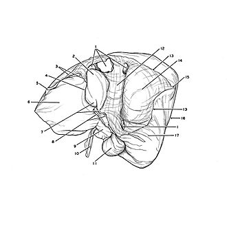
Posterosuperior aspect of liver
Image #144-6
KEYWORDS: Gallbladder, Liver, Pancreas, Spleen.
Creative Commons
Stanford holds the copyright to the David L. Bassett anatomical images and has assigned Creative Commons license Attribution-Share Alike 4.0 International to all of the images.
For additional information regarding use and permissions, please contact the Medical History Center.
Exploration of liver, gall bladder, pancreas, duodenum and spleen
Posterosuperior aspect of liver
- Hepatic veins
- Caudate lobe
- Right pointer: Cut edge of lesser omentum Left pointer: Left triangular ligament
- Fissure of ligamentum venosum
- Left triangular ligament of liver
- Left lobe of liver
- Upper pointer: Papillary process Lower pointer: Caudate process
- Upper pointer: Porta hepatis Lower pointer: Area of adhesion which obliterated normal peritoneal surface (normal situation depicted in drawing)
- Quadrate lobe
- Ligamentum teres
- Gallbladder
- Sulcus of vena cava
- Coronary ligament of liver
- Bare area of liver (note peritoneal reflection at margins of area (3, 13) which can be traced entirely around the region with the exception of an indistinct edge close to the porta hepatis (8, lower pointer)
- Right triangular ligament
- Right lobe of liver
- Renal impression (pointer indicates approximate midregion of this impression)


