Bassett Collection of Stereoscopic Images of Human Anatomy
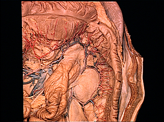
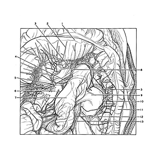
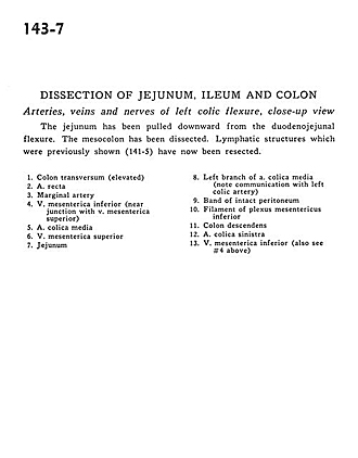
Dissection of jejunum, ileum and colon
Arteries, veins and nerves of left colic flexure, close-up view
Image #143-7
KEYWORDS: Large intestine, Peripheral nervous system, Small intestine, Vasculature.
Creative Commons
Stanford holds the copyright to the David L. Bassett anatomical images and has assigned Creative Commons license Attribution-Share Alike 4.0 International to all of the images.

For additional information regarding use and permissions, please contact the Medical History Center.
Dissection of jejunum, ileum and colon
Arteries, veins and nerves of left colic flexure, close-up view
The jejunum has been pulled downward from the duodenojejunal flexure. The mesocolon has been dissected. Lymphatic structures which were previously shown (141-5) have now been resected.
- Transverse colon (elevated)
- Straight artery
- Marginal artery
- Inferior mesenteric vein (near junction with superior mesenteric vein)
- Middle colic artery
- Superior mesenteric vein
- Jejunum
- Left branch of middle colic artery (note communication with left colic artery)
- Band of intact peritoneum
- Filament of inferior mesenteric plexus
- Descending colon
- Left colic artery
- Inferior mesenteric vein (also see #4 above)