Bassett Collection of Stereoscopic Images of Human Anatomy
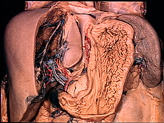
Dissection of stomach
Interior of stomach, anterior view
Image #142-6
KEYWORDS: Stomach.
Creative Commons
Stanford holds the copyright to the David L. Bassett anatomical images and has assigned Creative Commons license Attribution-Share Alike 4.0 International to all of the images.

For additional information regarding use and permissions, please contact the Medical History Center.
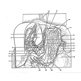

Dissection of stomach
Interior of stomach, anterior view
The anterior half of the stomach has been cut away.
- Margin of esophageal hiatus
- Caudate lobe of liver
- Proper hepatic artery (note hepatic plexus of nerves accompanying artery)
- Upper pointer: Portal vein Lower pointer: Common bile duct
- Body of pancreas
- Superior part of duodenum
- Descending part of duodenum
- Esophagus (left pointer, thoracic part; right pointer, abdominal part)
- Cardiac opening
- Fundus of stomach
- Spleen
- Muscular layer of stomach
- Gastric fold (lower part of this fold was removed in sectioning gastric wall)
- Canal of stomach
- Upper pointer: Muscular layer (circular fibers) Lower pointer: Submucosa of stomach
- Tail of pancreas
- Gastric fold
- Kidney (covered by renal fascia)
- Pyloric antrum
- Pyloric canal
- Upper pointer: Pyloric sphincter muscle Lower pointer: Pyloric opening
- Head of pancreas