Bassett Collection of Stereoscopic Images of Human Anatomy
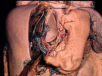
Dissection of stomach
Deep muscular layers of gastric wall, anterior view
Image #142-5
KEYWORDS: Muscles and tendons, Peripheral nervous system, Stomach, Vasculature.
Creative Commons
Stanford holds the copyright to the David L. Bassett anatomical images and has assigned Creative Commons license Attribution-Share Alike 4.0 International to all of the images.
For additional information regarding use and permissions, please contact the Medical History Center.
Dissection of stomach
Deep muscular layers of gastric wall, anterior view
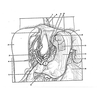
The outer, longitudinal layer of muscle has been partially removed from the cardiac part and from the anterior surface of the stomach. The underlying circular layer (13) has also been partially cut away to expose the innermost, obliquely-placed layer (12). The latter is an incomplete layer which arches over the cardiac notch and spreads downward in the anterior and posterior walls of the stomach.
- Hepatic vein
- Liver
- Upper pointer: Left gastric artery Lower pointer: Left gastric plexus
- Upper pointer: Common bile duct Lower pointer: Common hepatic artery
- Descending part of duodenum
- Pylorus (oblique fibers of pyloric sphincter exposed)
- Esophagus
- Left vagus nerve
- Margin of esophageal hiatus
- Muscular layer (longitudinal) note dissected area between pointers)
- Spleen
- Muscular layer (oblique fibers)
- Muscular layer (circular fibers)
- Body of stomach
- Tail of pancreas
- Kidney (covered by renal fascia)


