
Bassett Collection of Stereoscopic Images of Human Anatomy
Dissection of stomach
External muscular layer of gastric wall, posterior aspect
Image #142-4
KEYWORDS: Muscles and tendons, Peripheral nervous system, Stomach, Vasculature.
Creative Commons
Stanford holds the copyright to the David L. Bassett anatomical images and has assigned Creative Commons license Attribution-Share Alike 4.0 International to all of the images.

For additional information regarding use and permissions, please contact the Medical History Center.
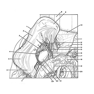

Dissection of stomach
External muscular layer of gastric wall, posterior aspect
The stomach has been rotated upward and to the right. The peritoneum which covered the posterior surface of the stomach has been removed to reveal the outer longitudinal layer of muscle.
- Major curvature of stomach
- Body of stomach (peritoneum removed to expose tunica muscularis)
- Lesser curvature of stomach
- Angular notch
- Pyloric antrum
- Pyloric canal
- Pylorus
- Thoracic aorta
- Fundus of stomach
- Diaphragm
- Spleen
- Left pointer: Esophagus (emerging through esophageal hiatus) Right pointer: Phrenic lymph node
- Phrenicolienal ligament (cut across)
- Esophageal branch of left phrenic artery
- Left gastric plexus
- Celiac lymph node
- Left gastric artery
- Pancreas
- Middle colic artery (lying approximately in position it occupied within mesotransverse colon)
- Jejunum (cut across)
- Caudate lobe
- Celiac lymph node
- Common hepatic artery