Bassett Collection of Stereoscopic Images of Human Anatomy
Exploration of peritoneal cavity
Retrocecal fossa; retrocecal appendix
Image #140-1
KEYWORDS:
Creative Commons
Stanford holds the copyright to the David L. Bassett anatomical images and has assigned Creative Commons license Attribution-Share Alike 4.0 International to all of the images.

For additional information regarding use and permissions, please contact the Medical History Center.
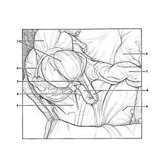

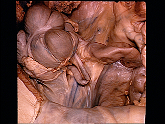
Exploration of peritoneal cavity
Retrocecal fossa; retrocecal appendix
Comparison of this specimen with the one shown in the preceding view (139-7) serves to demonstrate variations in the attachment of the cecum to the posterior abdominal wall. In the present example a cecal fold (5) forms one boundary of a deep retrocecal fossa into which the appendix extends.
- Cecum (elevated)
- Omental taenia coli
- Mesoappendix
- Retrocecal recess
- Cecal fold
- Mesenteries
- Ileum
- Vermiform appendix (sharply angulated; upper pointer indicates distal segment)
- Pelvic cavity