
Bassett Collection of Stereoscopic Images of Human Anatomy
Exploration of peritoneal cavity
Duodenojejunal junction; splenic flexure of colon
Image #139-2
KEYWORDS: Spleen.
Creative Commons
Stanford holds the copyright to the David L. Bassett anatomical images and has assigned Creative Commons license Attribution-Share Alike 4.0 International to all of the images.

For additional information regarding use and permissions, please contact the Medical History Center.
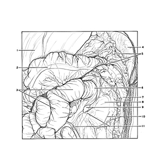

Exploration of peritoneal cavity
Duodenojejunal junction; splenic flexure of colon
The transverse colon has been elevated. Loops of the jejunum have been pulled to the right to expose the duodenojejunal junction. Peritoneal folds and recesses often associated with the duodenojejunal flexure are not well developed in this specimen. A small inferior duodenal reces (9)extends for a short distance downward beneath a broad, thin inferior duodenal fold of peritoneum.
- Liver
- Transverse colon (pointer on tenia coli)
- Jejunum
- Spleen
- Upper pointer: Greater omentum Lower pointer: Left colic flexure (splenic flexure)
- Duodenojejunal flexure
- Descending colon
- Position of kidney (covered by peritoneum and renal fascia)
- Upper pointer: Ascending part of duodenum Lower pointer: Inferior duodenal recess
- Branches of left colic artery (covered by peritoneum)
- Inferior duodenal fold