Bassett Collection of Stereoscopic Images of Human Anatomy
Exploration of peritoneal cavity
Upper abdominal organs in situ, diaphragm opened
Image #138-4
KEYWORDS: Overview.
Creative Commons
Stanford holds the copyright to the David L. Bassett anatomical images and has assigned Creative Commons license Attribution-Share Alike 4.0 International to all of the images.

For additional information regarding use and permissions, please contact the Medical History Center.
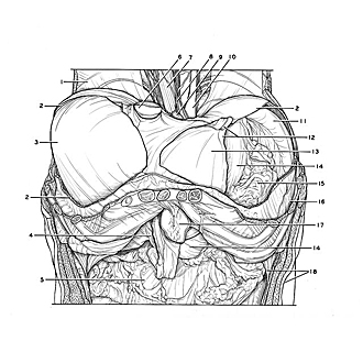
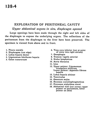
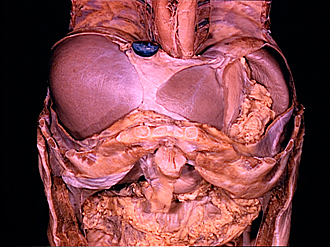
Exploration of peritoneal cavity
Upper abdominal organs in situ, diaphragm opened
Large openings have been made through the right and left sides of the diaphragm to expose the underlying organs. The reflections of the peritoneum from the diaphragm to the liver have been preserved. The specimen is viewed from above and in front.
- Costal pleura
- Diaphragm (cut edge)
- Right lobe of liver
- Falciform ligament of liver
- Transverse colon
- Inferior vena cava (cut at point of entry into right atrium)
- Esophagus
- Anterior vagal trunk
- Lymph node
- Thoracic aorta
- Spleen
- Upper pointer: Left triangular ligament Lower pointer: Left triangular ligament of liver
- Left lobe of liver
- Stomach
- Greater omentum
- Costodiaphragmatic recess
- Sternal xiphoid process
- Abdominal wall (cut across; upper pointer on peritoneum, lower pointer on skin)