
Bassett Collection of Stereoscopic Images of Human Anatomy
Dissection of female inguinal region
Inguinal canal (continued).
Image #137-4
KEYWORDS: Muscles and tendons, Overview.
Creative Commons
Stanford holds the copyright to the David L. Bassett anatomical images and has assigned Creative Commons license Attribution-Share Alike 4.0 International to all of the images.

For additional information regarding use and permissions, please contact the Medical History Center.

Dissection of female inguinal region
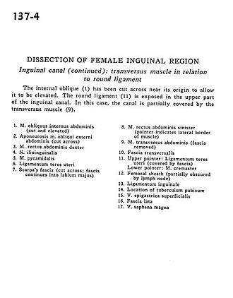
Inguinal canal (continued).
The internal oblique (1) has been cut across near its origin to allow it to be elevated. The round ligament *(11) is exposed in the upper part of the inguinal canal. In this case, the canal is partially covered by the transversus muscle (9).
- Internal oblique muscle (cut and elevated)
- Aponeurosis External oblique muscle (cut across)
- Right rectus abdominis muscle
- Ilioinguinal nerve
- Pyramidalis muscle
- Ligamentum teres (of uterus)
- Scarpa's fascia (cut across fascia continues into labium majus)
- Left rectus abdominis muscle (pointer indicates lateral border of muscle)
- Transversus abdominis muscle (fascia removed)
- Transversalis fascia
- Upper pointer: Ligamentum teres (of uterus) (covered by fascia) Lower pointer: Cremaster muscle
- Femoral sheath (partially obscured by lymph node)
- Inguinal ligament
- Location of pubic tubercle
- Superficial epigastric vein
- Fascia lata
- Great saphenous vein