Bassett Collection of Stereoscopic Images of Human Anatomy
Dissection of female inguinal region
Inguinal canal.
Image #137-3
KEYWORDS: Fascia, Muscles and tendons.
Creative Commons
Stanford holds the copyright to the David L. Bassett anatomical images and has assigned Creative Commons license Attribution-Share Alike 4.0 International to all of the images.

For additional information regarding use and permissions, please contact the Medical History Center.


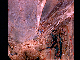
Dissection of female inguinal region
Inguinal canal.
The aponeurosis of the external oblique (4) has been split upward from the superficial inguinal ring in a direction parallel to the inguinal ligament. The underlying internal oblique muscle (5) appears between the reflected edges of the aponeurosis. Fascia has been removed from parts of the internal oblique and from the ilioinguinal nerve (6). Below the inferior border of the internal oblique the cremaster muscle and its associated cremasteric fascia (7) are visible lying in the lower part of the inguinal canal.
- Linea alba
- Upper pointer: Medial crus superficial inguinal ring Lower pointer: Ligamentum teres (of uterus)
- Location of pubic symphysis
- Aponeurosis external oblique muscle (divided)
- Internal oblique muscle
- Ilioinguinal nerve
- Upper pointer: Inferior border of internal oblique muscle Lower pointer: Cremasteric fascia (cremaster muscle visible through fascia)
- Lateral crus superficial inguinal ring
- Superficial inguinal lymph node
- External pudendal arteries
- Scarpa's fascia
- Labium majus