
Bassett Collection of Stereoscopic Images of Human Anatomy
Dissection of female inguinal region
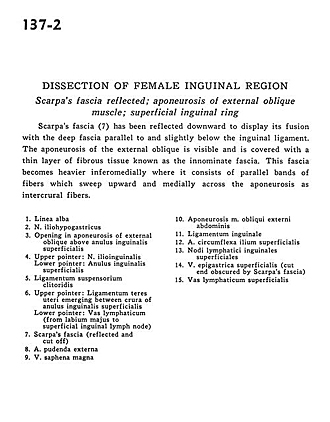
Scarpa's fascia reflected; aponeurosis of external oblique muscle; superficial inguinal ring
Image #137-2
KEYWORDS: Fascia, Muscles and tendons.
Creative Commons
Stanford holds the copyright to the David L. Bassett anatomical images and has assigned Creative Commons license Attribution-Share Alike 4.0 International to all of the images.

For additional information regarding use and permissions, please contact the Medical History Center.

Dissection of female inguinal region
Scarpa's fascia reflected; aponeurosis of external oblique muscle; superficial inguinal ring
Scarpa's fascia (7) has been reflected downward to display its fusion with the deep fascia parallel to and slightly below the inguinal ligament. The aponeurosis of the external oblique is visible and is covered with a thin layer of fibrous tissue known as the innominate fascia. This fascia becomes heavier inferomedially where it consists of parallel bands of fibres which sweep upward and medially across the aponeurosis as intercrural fibers.
- Linea alba
- Iliohypogastric nerve
- Opening in aponeurosis of external oblique above superficial inguinal ring
- Upper pointer: ilioinguinal nerve Lower pointer: Superficial inguinal ring
- Suspensory ligament of clitoris
- Upper pointer: Ligamentum teres (of uterus) emerging between crura of superficial inguinal ring Lower pointer: Lymph vessel (from labium majus to superficial inguinal lymph node)
- Scarpa's fascia (reflected and cut off)
- External pudendal artery
- Great saphenous vein
- Aponeurosis external oblique muscle
- Inguinal ligament
- Superficial circumflex iliac artery
- Superficial inguinal lymph node
- Superficial epigastric vein cut end obscured by Scarpa's fascia)
- Superficial lymph vessel