Bassett Collection of Stereoscopic Images of Human Anatomy
Dissection of anterolateral abdominal wall
Composition of rectus sheath in lower abdominal wall
Image #135-1
KEYWORDS: Muscles and tendons.
Creative Commons
Stanford holds the copyright to the David L. Bassett anatomical images and has assigned Creative Commons license Attribution-Share Alike 4.0 International to all of the images.

For additional information regarding use and permissions, please contact the Medical History Center.

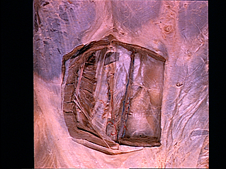
Dissection of anterolateral abdominal wall
Composition of rectus sheath in lower abdominal wall
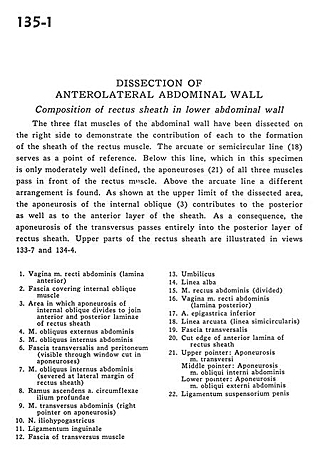
The three flat muscles of the abdominal wall have been dissected on the right side to demonstrate the contribution of each of the formation of the sheath of the rectus muscle. The arcuate or semicircular line (18) serves as a point of reference. Below this line, which in this specimen is only moderately well defined, the aponeuroses (21) of all three muscles pass in front of the rectus muscle. Above the arcuate line a different arrangement was found. As shown at the upper limit of the dissected area, the aponeurosis of the internal oblique (3) contributes to the posterior as well as to the anterior layer of the sheath. As a consequence, the aponeurosis of the transversus pass entirely into the posterior layer of rectus sheath. Upper parts of the rectus sheath are illustrated in views 133-7 and 134-4.
- Sheath of rectus abdominis muscle (anterior layer)
- Fascia covering internal oblique muscle
- Area in which aponeurosis of internal oblique divides to join anterior and posterior layers of rectus sheath
- External oblique muscle
- Internal oblique muscle
- Transversalis fascia and peritoneum (visible through window cut in aponeuroses)
- Internal oblique muscle (severed at lateral margin of rectus sheath)
- Ascending branch deep circumflex iliac artery
- Transversus abdominis muscle (right pointer on aponeurosis)
- Iliohypogastric nerve
- Inguinal ligament
- Fascia of transversus muscle
- Umbilicus
- Linea alba
- Rectus abdominis muscle (divided)
- Sheath of rectus abdominis muscle (posterior layer)
- Inferior epigastric artery
- Arcuate line (semicircular line)
- Transversalis fascia
- Cut edge of anterior layer of rectus sheath:
- Upper pointer: Aponeurosis transversus muscle Middle pointer: Aponeurosis of Internal oblique muscle Lower pointer: Aponeurosis external oblique muscle
- Suspensory ligament of the penis