Bassett Collection of Stereoscopic Images of Human Anatomy
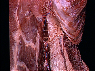
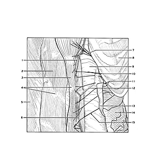
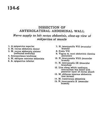
Dissection of anterolateral abdominal wall
Nerve supply to left rectus abdominis, close-up view of midportion of muscle
Image #134-6
KEYWORDS: Muscles and tendons, Peripheral nervous system.
Creative Commons
Stanford holds the copyright to the David L. Bassett anatomical images and has assigned Creative Commons license Attribution-Share Alike 4.0 International to all of the images.

For additional information regarding use and permissions, please contact the Medical History Center.
Dissection of anterolateral abdominal wall
Nerve supply to left rectus abdominis, close-up view of midportion of muscle
- Superior epigastric artery
- Right rectus abdominis muscle
- Left rectus abdominis muscle (reflected medially)
- Tendinous inscriptions
- External oblique muscle
- Inferior epigastric artery
- Intercostal nerve VII (muscular branch)
- Rib VII
- Sheath of rectus abdominis muscle (posterior layer)
- Intercostal nerve VIII (muscular branch)
- Intercostal nerve IX (muscular branches)
- Line along which tendinous intersection was fused with posterior layer of rectus sheath
- Internal oblique muscle (cut across)
- Transversus abdominis muscle
- Intercostal nerve X (muscular branch)