Bassett Collection of Stereoscopic Images of Human Anatomy
Dissection of anterolateral abdominal wall
Internal oblique muscle, left lateral view
Image #134-1
KEYWORDS: Muscles and tendons.
Creative Commons
Stanford holds the copyright to the David L. Bassett anatomical images and has assigned Creative Commons license Attribution-Share Alike 4.0 International to all of the images.

For additional information regarding use and permissions, please contact the Medical History Center.
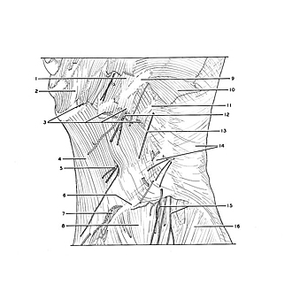
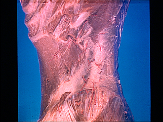
Dissection of anterolateral abdominal wall
Internal oblique muscle, left lateral view
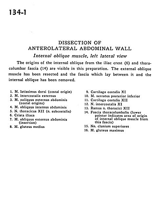
The origins of the internal oblique from the iliac crest(6) and thoracolumbar fascia (14) are visible in this preparation. The external oblique muscle has been resected and the fascia which lay between it and the internal oblique has been removed.
- Latissimus dorsi muscle (costal origin)
- External intercostal muscle
- External oblique muscle (costal origins)
- Internal oblique muscle
- Thoracic nerve XII (subcostal nerve)
- Iliac crest
- External oblique muscle (insertion)
- Gluteus medius muscle
- Costal cartilage XI
- Serratus posterior inferior muscle
- Costal cartilage XII
- Intercostal nerve XI
- Branch of thoracic nerve XII
- Thoracolumbar fascia (lower pointer indicates area of origin of internal oblique muscle from this fascia)
- Superior cluneal nerves
- Gluteus maximus muscle