
Bassett Collection of Stereoscopic Images of Human Anatomy
Dissection of anterolateral abdominal wall
Nerve supply to external oblique muscle (continued)
Image #133-4
KEYWORDS: Muscles and tendons, Peripheral nervous system.
Creative Commons
Stanford holds the copyright to the David L. Bassett anatomical images and has assigned Creative Commons license Attribution-Share Alike 4.0 International to all of the images.

For additional information regarding use and permissions, please contact the Medical History Center.
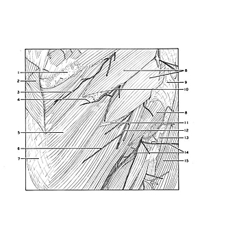

Dissection of anterolateral abdominal wall
Nerve supply to external oblique muscle (continued)
The upper fascicles of the left external oblique muscle are shown in this close-up photograph. Nerves (3,4,6,14) which supply the muscle originate from lateral cutaneous branches of the intercostal nerves and enter the muscle from its external surface.
- Rib VI (covered by periosteum)
- Pectoralis major muscle (abdominal part, reflected)
- Muscular branch of lateral cutaneous branch thoracie nerve VI
- Muscular branch of lateral cutaneous branch thoracic nerve VII
- External oblique muscle
- Muscular branch of lateral cutaneous branch thoracic nerve VIII
- Aponeurosis external oblique muscle
- Serratus anterior muscle
- Fascia of serratus anterior muscle
- Lateral cutaneous branch thoracic nerve VII
- Rib VIII
- Lateral cutaneous branch thoracic nerve VIII
- Rib X
- Upper pointer: Lateral cutaneous branch thoracic nerve IX Lower pointer: Muscular branch 15. Latissimus dorsi muscle