
Bassett Collection of Stereoscopic Images of Human Anatomy
Dissection of diaphragm
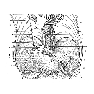
Diaphragm viewed from above, pleura removed
Image #130-1
KEYWORDS: Diaphragm, Lung, Muscles and tendons, Pericardial sac.
Creative Commons
Stanford holds the copyright to the David L. Bassett anatomical images and has assigned Creative Commons license Attribution-Share Alike 4.0 International to all of the images.

For additional information regarding use and permissions, please contact the Medical History Center.

Dissection of diaphragm
Diaphragm viewed from above, pleura removed
The lungs have been removed and the tracheobronchial tree has been pulled posteriorly to provide an unobstructed view of the diaphragm. A narrow band of pericardium has been preserved along the margin of its diaphragmatic attachment. Lymphatic structures have been dissected on the upper surface of the diaphragm.
- Costal pleura
- Ascending aorta
- Esophagus
- Phrenic nerve right
- Central tendon of diaphragm
- Inferior vena cava
- Lymph vessel
- Pericardium
- Musculophrenic artery
- Left main bronchus (retracted)
- Rib VII (cut off)
- Posterior mediastinal lymph node
- Left vagus nerve
- Phrenicoesophageal membrane
- Phrenic nerve left (cut off)
- Musculophrenic artery