Bassett Collection of Stereoscopic Images of Human Anatomy
Dissection of diaphragm
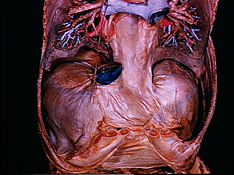
Diaphragm viewed from above, pleura and diaphragmatic part of pericardium intact
Image #129-7
KEYWORDS: Diaphragm, Lung, Muscles and tendons, Pericardial sac, Peripheral nervous system, Pleura.
Creative Commons
Stanford holds the copyright to the David L. Bassett anatomical images and has assigned Creative Commons license Attribution-Share Alike 4.0 International to all of the images.

For additional information regarding use and permissions, please contact the Medical History Center.


Dissection of diaphragm
Diaphragm viewed from above, pleura and diaphragmatic part of pericardium intact
The heart and the anterior part of the pericardium have been removed. The upper lobes of the lungs have been cut away and the lower lobes have been dissected. The right phrenic nerve is stretched upward approximately in its normal course, while the left nerve has been cut off.
- Left atrium
- Left inferior pulmonary vein (cut off at entrance into left atrium)
- Pericardium
- Inferior lobe right lung
- Right phrenic nerve
- Inferior vena cava (cut off at entrance into right atrium)
- Diaphragm (pointers on right and left divisions of central tendon)
- Diaphragm (muscular portion)
- Costal cartilage VII
- Lower lobe left lung
- Pulmonary ligament
- Phrenic nerve left
- Xiphoid process (cut off)