Bassett Collection of Stereoscopic Images of Human Anatomy
Dissection of mediastinum and paravertebral structures
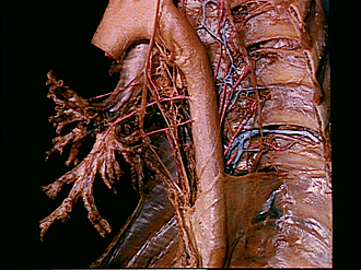
Bronchial and esophageal arteries viewed from left side
Image #127-7
KEYWORDS: Bones joints cartilage, Left lung, Lung, Lymphatics, Mediastinum, Right lung, Vasculature, Vertebral column.
Creative Commons
Stanford holds the copyright to the David L. Bassett anatomical images and has assigned Creative Commons license Attribution-Share Alike 4.0 International to all of the images.

For additional information regarding use and permissions, please contact the Medical History Center.


Dissection of mediastinum and paravertebral structures
Bronchial and esophageal arteries viewed from left side
The tracheobronchial tree has been retracted anteriorly in order to stretch the bronchial arteries (15). Delicate lymphatic tissue (19) has been partially preserved in the intercostal spaces and along the vertebral bodies.
- Aortic arch
- Trachea
- Left main bronchus
- Bronchial branch of vagus nerve
- Left upper lobe bronchus
- Bronchopulmonary lymph node (partially removed)
- Left lower lobe bronchus
- Inferior tracheobronchial lymph node
- Thoracic duct (dark amber color)
- Esophagus
- Lymph vessel
- Left vagus nerve
- Recurrent laryngeal nerve
- Ligamentum arteriosum (cut off)
- Upper and middle pointers: Bronchial branches thoracic aorta Lower pointer: Esophageal branch thoracic aorta
- Rib V
- Sympathetic trunk
- Intercostal nerve V
- Upper pointer: Intercostal lymph node Lower pointer: Lymph vessel (a considerable plexus of lymphatic vessels is visible along the sides of the vertebrae and heads of ribs and in the intercostal spaces. Only in this area of the drawing is this plexus included and labeled.)
- Thoracic ganglion
- Endothoracic fascia
- Intercostal artery VI
- Accessory hemiazygos vein
- Esophageal branch of sympathetic trunk
- Thoracic aorta
- Costal pleura
- Pulmonary ligament (cut)