Bassett Collection of Stereoscopic Images of Human Anatomy
Dissection of mediastinum and paravertebral structures
Superior mediastinum.
Image #127-4
KEYWORDS: Left lung, Lung, Mediastinum, Pericardial sac, Peripheral nervous system, Vasculature.
Creative Commons
Stanford holds the copyright to the David L. Bassett anatomical images and has assigned Creative Commons license Attribution-Share Alike 4.0 International to all of the images.

For additional information regarding use and permissions, please contact the Medical History Center.


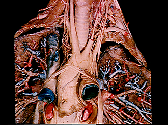
Dissection of mediastinum and paravertebral structures
Superior mediastinum.
The lymph nodes and vessels which were shown in the previous view have been removed from the dissection.
- Vagus nerve right
- Inferior cervical cardiac vein
- Inferior cardiac branch of vagus nerve
- Right brachiocephalic vein
- Brachiocephalic trunk
- Posterior mediastinal lymph node
- Filaments of cardiac plexus (two pointers encompass plexus)
- Superior vena cava (cut across)
- Right coronary artery (cut off)
- Right superior pulmonary vein (at entrance into left atrium)
- Left pointer: Trachea Right pointer: Esophagus
- Stellate ganglion
- Middle cervical cardiac nerve
- Thoracic duct (cut off)
- Superior cardiac branch vagus nerve
- Vagus nerve left
- Left subclavian artery
- Recurrent laryngeal nerve
- Pericardium
- Pulmonary trunk
- Cardiac ganglion
- Left coronary artery