Bassett Collection of Stereoscopic Images of Human Anatomy
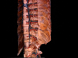
Views of segmental bronchi, azygos system of viens, sympathetic trunks and costovertebral joints
Sympathetic trunk, rami communicantes and splanchnic nerves in lower thoracic region, left anterolateral view
Image #126A-6
KEYWORDS: Bones joints cartilage, Peripheral nervous system, Vertebral column.
Creative Commons
Stanford holds the copyright to the David L. Bassett anatomical images and has assigned Creative Commons license Attribution-Share Alike 4.0 International to all of the images.

For additional information regarding use and permissions, please contact the Medical History Center.
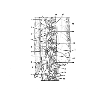

Views of segmental bronchi, azygos system of viens, sympathetic trunks and costovertebral joints
Sympathetic trunk, rami communicantes and splanchnic nerves in lower thoracic region, left anterolateral view
The specimen shown in the previous view has been turned to expose its left anterolateral aspect in this close-up view of the lower thoracic and upper lumbar part of the spine.
- Accessory hemiazygos vein
- Azygos vein
- Upper pointer: Body of vertebra Th. VIII (pointer on anterior longitudinal ligament)
- Vein draining body of vertebra
- Posterior intercostal arteries IX-X
- Major splanchnic nerve (lower pointer on splanchnic ganglion)
- Minor splanchnic nerve
- Smallest splanchnic nerve
- Diaphragm (cut off)
- Lumbar part of diaphragm (upper pointer: left crus; lower pointer: right crus)
- Left pointer: Hemiazygos vein Right pointer: Posterior intercostal vein VIII
- Rib VIII
- Costal pleura
- Sympathetic trunk (lower pointer on sympathetic trunk ganglion)
- Communicating branches
- Intercostal nerve XI
- Head of rib XII
- Subcostal nerve (note large communicating branch)
- Upper pointer: Lumbar quadratic muscle Lower pointer: Psoas major muscle
- Ascending lumbar vein
- Transverse process of vertebra L. II
- Lumbar nerve I (passing downward to join lumbar plexus)
- [Legend above restored translation from Latin]