Bassett Collection of Stereoscopic Images of Human Anatomy
Dissection of mediastinum and paravertebral structures
Mediastinal contents viewed from left side
Image #126-6
KEYWORDS: Mediastinum, Pericardial sac, Pleura.
Creative Commons
Stanford holds the copyright to the David L. Bassett anatomical images and has assigned Creative Commons license Attribution-Share Alike 4.0 International to all of the images.

For additional information regarding use and permissions, please contact the Medical History Center.
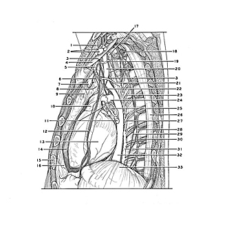

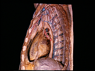
Dissection of mediastinum and paravertebral structures
Mediastinal contents viewed from left side
The pleura has been removed from the left side of the mediastinum and from the paravertebral area. The pericardial cavity has been opened.
- Subclavius muscle (cut off)
- Left subclavian artery and vein
- Thoracic duct
- Rib I (periosteum removed)
- Internal thoracic artery and vein
- Pericardiacophrenic artery
- Left brachiocephalic vein (in background)
- Aortic arch (covered by fibrous tissue)
- Thymus (covered by fascia)
- Pulmonary trunk
- Left pulmonary veins (filled with red latex)
- Phrenic nerve left (band of pericardium preserved along nerve)
- Left ventricle
- Pericardial cavity
- Costal cartilage V
- Pericardium (opened)
- Longus colli muscle
- Intercostal nerve II
- Supreme intercostal vein
- Upper pointer: Left subclavian artery Lower pointer: Esophagus
- Left superior intercostal vein
- Vagus nerve left
- Posterior intercostal artery IV
- Left pulmonary artery (filled with blue latex)
- Left main bronchus
- Costal pleura
- Accessory hemiazygos vein
- Thoracic ganglion
- Greater splanchnic nerve
- Sympathetic trunk
- Hemiazygos vein
- Thoracic aorta
- Diaphragm