Bassett Collection of Stereoscopic Images of Human Anatomy
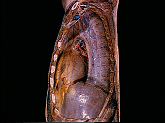
Dissection of mediastinum and paravertebral structures
Mediastinal pleura viewed from left side
Image #126-5
KEYWORDS: Mediastinum, Pleura.
Creative Commons
Stanford holds the copyright to the David L. Bassett anatomical images and has assigned Creative Commons license Attribution-Share Alike 4.0 International to all of the images.

For additional information regarding use and permissions, please contact the Medical History Center.


Dissection of mediastinum and paravertebral structures
Mediastinal pleura viewed from left side
The upper limb has been detached. The left pleural cavity has been opened and the lung has been excised.
- Clavicle (cut across)
- Brachial plexus
- Subclavian artery and vein
- Pectoralis major muscle (clavicular part, cut across)
- Rib I (intact)
- Internal thoracic artery
- Line of reflection of mediastinal pleura onto lung
- Branches left upper pulmonary vein
- Pectoralis major muscle (sternal part, cut off)
- Costal cartilage ill (cut across)
- Transversus thoracis muscle (cut across)
- Costomediastinal recess
- Mediastinal pleura (pointer indicates location of phrenic nerve in its course across left border of heart and pericardium)
- Lobule of fat
- Costodiaphragmatic recess
- Rib III (cut off)
- Cupula pleurae
- Aortic arch (covered by pleura)
- Left pulmonary artery (filled with blue latex)
- Left main bronchus
- Left inferior pulmonary vein
- Thoracic aorta (covered by pleura)
- Pulmonary ligament
- Costal pleura
- Diaphragm
- Rib X (periosteum removed)