
Bassett Collection of Stereoscopic Images of Human Anatomy
Dissection of mediastinum and paravertebral structures
Mediastinal contents viewed from right side
Image #126-4
KEYWORDS: Mediastinum, Pericardial sac, Pleura.
Creative Commons
Stanford holds the copyright to the David L. Bassett anatomical images and has assigned Creative Commons license Attribution-Share Alike 4.0 International to all of the images.

For additional information regarding use and permissions, please contact the Medical History Center.
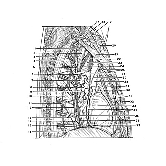
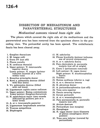
Dissection of mediastinum and paravertebral structures
Mediastinal contents viewed from right side
The pleura which covered the right side of the mediastinum and the paravertebral area has been removed from the specimen shown in the preceding view. The pericardial cavity has been opened. The endothoracic fascia has been cleared away.
- Thoracic ganglion
- Longus colli muscle
- Rib IV (cut off)
- Costal pleura
- Cardiac thoracic nerve
- Upper pointer: Right superior intercostal vein Lower pointer: Azygos vein (pointer indicates location of a valve within vein)
- Right main bronchus
- Branch right pulmonary artery (filled with blue latex)
- Right pulmonary veins (filled with red latex)
- Ligamentum capitis costae radiatum
- Upper pointer: Ramus communicans Lower pointer: Thoracic ganglion
- Greater splanchnic nerve (two strands indicated by pointers join at a lower level)
- Posterior intercostal artery and vein
- Anterior longitudinal ligament
- Sympathetic trunk
- Diaphragm
- Subclavius muscle
- Brachial plexus (pointer indicates one of several components)
- Right subclavian artery and vein
- Clavicular part pectoralis major muscle (cut off at origin)
- Rib I (periosteum removed)
- Left pointer: Right vagus nerve Right pointer: Right brachiocephalic vein
- Trachea
- Inferior cardiac branch of vagus nerve
- Internal thoracic artery and vein
- Right phrenic nerve
- Pericardiacophrenic artery (cut off)
- Superior vena cava
- Fascia covering thymus
- Pericardial cavity
- Musculophrenic artery and vein
- Sternocostal part pectoralis major muscle (cut off)
- Right atrium
- Pericardium (cut margins)
- Esophagus
- Vagus nerve right (note filaments of esophageal plexus above pointer)
- Inferior vena cava (covered by pericardium)