Bassett Collection of Stereoscopic Images of Human Anatomy
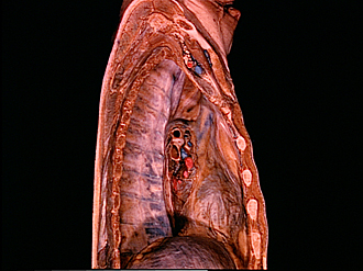
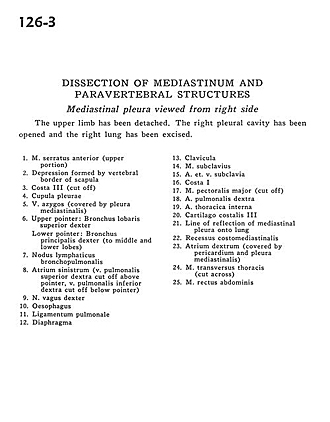
Dissection of mediastinum and paravertebral structures
Mediastinal pleura viewed from right side
Image #126-3
KEYWORDS: Mediastinum, Pleura.
Creative Commons
Stanford holds the copyright to the David L. Bassett anatomical images and has assigned Creative Commons license Attribution-Share Alike 4.0 International to all of the images.

For additional information regarding use and permissions, please contact the Medical History Center.

Dissection of mediastinum and paravertebral structures
Mediastinal pleura viewed from right side
The upper limb has been detached. The right pleural cavity has been opened and the right lung has been excised.
- Serratus anterior muscle (upper portion)
- Depression formed by vertebral border of scapula
- Rib III (cut off)
- Cupula pleurae
- Azygos vein (covered by mediastinal pleura)
- Upper pointer: Bronchus of upper right lobe Lower pointer: Right main bronchus (to middle and lower lobes)
- Bronchopulmonary lymph node
- Left atrium (right superior pulmonary vein cut off above pointer, right inferior pulmonary vein cut off below pointer)
- Right vagus nerve
- Esophagus
- Pulmonary ligament
- Diaphragm
- Clavicle
- Subclavius muscle
- Subclavian artery and vein
- Rib I
- Pectoralis major muscle (cut off)
- Right pulmonary artery
- Internal thoracic artery
- Costal cartilage III
- Line of reflection of mediastinal pleura onto lung
- Costomediastinal recess
- Right atrium (covered by pericardium and mediastinal pleura)
- Transversus thoracis muscle (cut across)
- Rectus abdominis muscle