
Bassett Collection of Stereoscopic Images of Human Anatomy
Dissection of lungs in situ
Left lung.
Image #126-2
KEYWORDS: Left lung, Lung, Vasculature.
Creative Commons
Stanford holds the copyright to the David L. Bassett anatomical images and has assigned Creative Commons license Attribution-Share Alike 4.0 International to all of the images.

For additional information regarding use and permissions, please contact the Medical History Center.
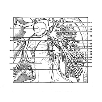

Dissection of lungs in situ
Left lung.
Branches of the pulmonary artery and vein have been removed to demonstrate the segmental bronchi (12, 13, 16, 19, 20, 22) of the lower lobe. The view is directed medially.
- Pulmonary trunk
- Left main bronchus
- Upper pointer: Superior lingular bronchus (in foreground) Lower pointer: Inferior lingular bronchus (in foreground)
- Left inferior pulmonary vein
- Posterior mediastinal lymph node
- Pericardium
- Pulmonary ligament
- Left pulmonary artery (cut off)
- Posterior apical segmental bronchus
- Interlobar surface lower lobe
- Anterior segmental bronchus
- Superior segmental bronchus (pointer on superior branch)
- Superior segmental bronchus (pointer on lateral branch)
- Posterior and lateral basilar branches left pulmonary artery
- Upper pointer: Left lower lobe bronchus Lower pointer: Bronchial branch of aorta
- Anterior basal segmental bronchus
- Lateral branch of no. 16
- Basal branch of no.16
- Medial basal segmental bronchus
- Lateral basal segmental bronchus
- Upper pointer: Anteromedial branch of no. 19 Lower pointer: Anterolateral branch of no. 19
- Posterior basal segmental bronchus (division of this bronchus into laterobasal and mediobasal branches is visible below pointer)