
Bassett Collection of Stereoscopic Images of Human Anatomy
Creative Commons
Stanford holds the copyright to the David L. Bassett anatomical images and has assigned Creative Commons license Attribution-Share Alike 4.0 International to all of the images.

For additional information regarding use and permissions, please contact the Medical History Center.
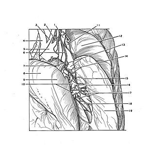
Dissection of lungs in situ
Left lung.
The left upper lobe has been retracted anteriorly and toward the right. The pleura in the depths of the oblique fissure has been cut to expose the interlobar parts of the left pulmonary artery, the pulmonary plexus of nerves and bronchopulmonary lymph nodes and vessels. Unlike the situation in the right lung (124-7), there is no area of fusion of upper and lower lobes on the left.
- Left subclavian artery
- Left vertebral artery (unusual origin from aortic arch)
- Left common carotid artery
- Brachiocephalic trunk
- Aortic arch
- Left vagus nerve
- Ligamentum arteriosum
- Upper lobe (retracted)
- Interlobar surface upper lobe
- Posterior branches left pulmonary artery (entering upper lobe)
- Posterior mediastinal lymph node
- Costal pleura (pointer on third rib)
- Upper pointer: Recurrent laryngeal nerve Lower pointer: Thoracic aorta
- Upper pointer: Vagus nerve Lower pointer: Branch entering pulmonary plexus
- Line of reflection of pleura between lobes of lung
- Left pulmonary artery
- Bronchopulmonary lymph nodes
- Left lower lobe bronchus
- Interlobar surface lower lobe