Bassett Collection of Stereoscopic Images of Human Anatomy
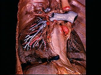
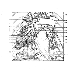
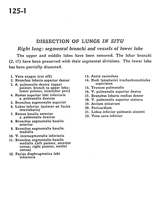
Dissection of lungs in situ
Right lung.
Image #125-1
KEYWORDS: Lung, Right lung, Vasculature.
Creative Commons
Stanford holds the copyright to the David L. Bassett anatomical images and has assigned Creative Commons license Attribution-Share Alike 4.0 International to all of the images.

For additional information regarding use and permissions, please contact the Medical History Center.
Dissection of lungs in situ
Right lung.
The upper and middle lobes have been removed. The lobar bronchi (2, 17) have been preserved with their segmental divisions. The lower lobe has been partially dissected.
- Azygos vein (cut off)
- Bronchus of upper right lobe
- Right pulmonary artery (upper pointer, branch to upper lobe lower pointer, interlobar part)
- Superior branch lower lobe right pulmonary artery
- Superior segmental bronchus
- Lower lobe (pointer on interlobar surface)
- Basal branch anterior right pulmonary artery
- Anterior basal segmental bronchus
- Medial basal segmental bronchus
- Inferior intersegmental vein
- Medial basal segmental bronchus (left pointer, anterior branch; right pointer, medial branch)
- Diaphragmatic surface lower lobe
- Ascending aorta
- Superior tracheobronchial lymph nodes
- Pulmonary trunk
- Right superior pulmonary vein
- Bronchus of middle right lobe
- Left superior pulmonary vein
- Left atrium
- Pericardium
- Lower lobe left lung
- Inferior vena cava