Bassett Collection of Stereoscopic Images of Human Anatomy
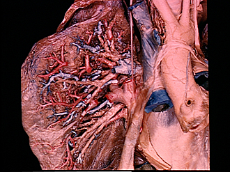
Dissection of lungs in situ
Right lung.
Image #124-5
KEYWORDS: Lung, Right lung, Vasculature.
Creative Commons
Stanford holds the copyright to the David L. Bassett anatomical images and has assigned Creative Commons license Attribution-Share Alike 4.0 International to all of the images.

For additional information regarding use and permissions, please contact the Medical History Center.
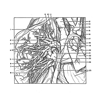

Dissection of lungs in situ
Right lung.
The dissection has been carried more deeply into the lung. Various branches of the pulmonary vessels have been divided or partially removed to permit better visualization of the bronchial distribution.
- Branches of apical branch right pulmonary artery (cut off refer to no. in previous view)
- Apical segmental bronchus (note division above pointer into apical and anterior branches)
- Anterior segmental bronchus (note division near pointer into anterior and posterior branches)
- Anterior branch right pulmonary artery (cut off refer to no. 5 in previous view)
- Intersegmental vein
- Lateral segmental bronchus middle lobe (anterior branch)
- Medial segmental bronchus of middle lobe (note division into superior and inferior branches)
- Vein of middle lobe (note division into medial and lateral branches)
- Superior intersegmental vein
- Apical branches of apical branch right pulmonary artery
- Posterior branch right pulmonary artery
- Right brachiocephalic vein
- Left common carotid artery
- Right phrenic nerve
- Left brachiocephalic vein (cut off)
- Left vagus nerve
- Filament of pulmonary plexus
- Superior vena cava
- Right pulmonary artery (entering upper lobe)
- Cardiac plexus
- Bronchopulmonary lymph node
- Ascending aorta
- Right superior pulmonary vein
- Branch of right pulmonary artery (to middle lobe)
- Pericardium