Bassett Collection of Stereoscopic Images of Human Anatomy
Dissection of lungs in situ
Right lung.
Image #124-4
KEYWORDS: Lung, Right lung, Vasculature.
Creative Commons
Stanford holds the copyright to the David L. Bassett anatomical images and has assigned Creative Commons license Attribution-Share Alike 4.0 International to all of the images.

For additional information regarding use and permissions, please contact the Medical History Center.

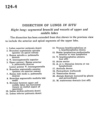
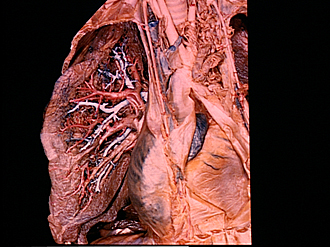
Dissection of lungs in situ
Right lung.
The dissection has been extended from that shown in the previous view to include the anterior and apical segments of the upper lobe.
- Upper lobe right lung
- Apical segmental bronchus (pointer on apical branch)
- Apical branch right pulmonary artery
- Superior intersegmental vein
- Upper pointer: Anterior branch right pulmonary artery Lower pointer Intersegmental vein
- Anterior segmental bronchus (pointer on anterior branch)
- Branch to middle lobe right pulmonary artery
- Medial segmental bronchus of middle lobe
- Division between upper and middle lobes (no horizontal fissure on medial aspect of specimen)
- Middle lobe right lung
- Inferior lobe right lung
- Brachiocephalic trunk and right brachiocephalic vein
- Anterior mediastinal lymph node and lymph vessel
- Left brachiocephalic vein (cut off)
- Aortic arch
- Internal thoracic artery and vein and lymph vessel
- Right auricle
- Pericardium (reflected)
- Right ventricle
- Right atrium (covered by pleura and pericardium)
- Transversus thoracis muscle (cut off)