
Bassett Collection of Stereoscopic Images of Human Anatomy
Dissection of lungs in situ
Bronchogram, anteroposterior view
Image #123-6
KEYWORDS: Left lung, Lung, Right lung.
Creative Commons
Stanford holds the copyright to the David L. Bassett anatomical images and has assigned Creative Commons license Attribution-Share Alike 4.0 International to all of the images.

For additional information regarding use and permissions, please contact the Medical History Center.
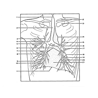

Dissection of lungs in situ
Bronchogram, anteroposterior view
This film of a living subject was made after the bronchial tree had been partially filled with iodized oil. The air passages are distinguished in some places by their content of air (e.g. 1, 2) and elsewhere by their content of radio-opaque material either alone or in combination with air. The manner of branching of the bronchi within both lower lobes is partially obscured by overlapping shadows. Details of the bronchial distribution in these lobes are shown in the lobar dissections which follow.
- Conus elasticus (air-filled)
- Trachea (air-filled)
- Apical segmental bronchus of upper lobe
- Posterior segmental bronchus of upper lobe
- Anterior segmental bronchus of upper lobe
- Superior segmental bronchus of lower lobe
- Upper pointer: Lateral segmental bronchus of middle lobe Lower pointer: Medial segmental bronchus of middle lobe
- Left pointer: Anterior basal segmental bronchus Right pointer: Medial basal segmental bronchus
- Lateral basal segmental bronchus
- Posterior basal segmental bronchus
- Diaphragm
- Rib I
- Clavicle
- Left border of aortic arch
- Upper pointer: Posterior apical segmental bronchus Lower pointer: Anterior segmental bronchus
- Main bronchi
- Upper pointer: Superior lingular bronchus Lower pointer: Inferior lingular bronchus
- Medial basal segmental bronchus
- Lateral basal segmental bronchus
- Anterior basal segmental bronchus
- Upper pointer: Posterior basal segmental bronchus Lower pointer: Heart