Bassett Collection of Stereoscopic Images of Human Anatomy
Detailed dissection of heart
Tricuspid valve viewed from below
Image #121-6
KEYWORDS: Heart, Right heart.
Creative Commons
Stanford holds the copyright to the David L. Bassett anatomical images and has assigned Creative Commons license Attribution-Share Alike 4.0 International to all of the images.
For additional information regarding use and permissions, please contact the Medical History Center.
Detailed dissection of heart
Tricuspid valve viewed from below
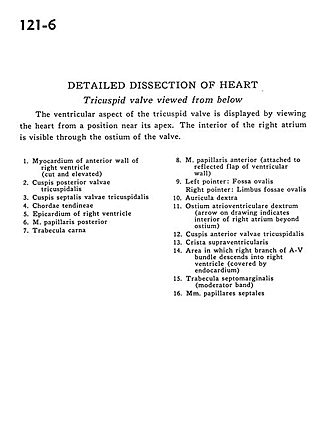
The ventricular aspect of the tricuspid valve is displayed by viewing the heart from a position near its apex. The interior of the right atrium is visible through the ostium of the valve.
- Myocardium of anterior wall of right ventricle (cut and elevated)
- Posterior cusp tricuspid valve
- Septal (medial) cusp of tricuspid valve
- Chordae tendineae
- Epicardium of right ventricle
- Posterior papillary muscle
- Trabeculae carnae
- Anterior papillary muscle (attached to reflected flap of ventricular wall)
- Left pointer: Fossa ovalis Right pointer: Border of fossa ovalis
- Right auricle
- Right atrioventricular opening (arrow on drawing indicates interior of right atrium beyond opening)
- Anterior cusp tricuspid valve
- Supraventricular crest
- Area in which right branch of A-V bundle descends into right ventricle (covered by endocardium)
- Septomarginal trabecula (moderator band)
- Septal papillary muscles


