
Bassett Collection of Stereoscopic Images of Human Anatomy
Detailed dissection of heart
Interior of right ventricle, anterior view
Image #121-4
KEYWORDS: Heart, Right heart.
Creative Commons
Stanford holds the copyright to the David L. Bassett anatomical images and has assigned Creative Commons license Attribution-Share Alike 4.0 International to all of the images.

For additional information regarding use and permissions, please contact the Medical History Center.

Detailed dissection of heart
Interior of right ventricle, anterior view
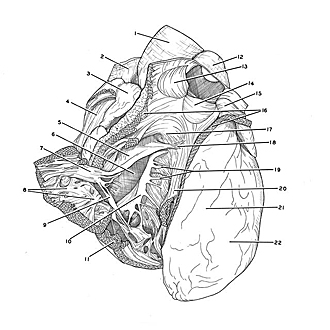
The right ventricle, conus arteriosus and pulmonary trunk have been opened. The part of the ventricular wall which gives attachment to the anterior papillary muscle (7) has been reflected. The right atrium remains open from a previous dissection.
- Ascending aorta
- Superior vena cava
- Right auricle
- Right atrium (opened)
- Anterior cusp tricuspid valve
- Right atrioventricular opening
- Anterior papillary muscle
- Trabeculae carnae
- Posterior cusp tricuspid valve
- Septal (medial) cusp of tricuspid valve
- Cut edge of myocardium of right ventricle
- Pulmonary trunk
- Right semilunar cusp
- Semilunar valves
- Valve of left pulmonary trunk Anterior semilunar cusp
- Conus arteriosus (cut margins retracted)
- Supraventricular crest
- Septal papillary muscle
- Chordae tendineae
- Septomarginal trabecula (moderator band)
- Position of anterior interventricular sulcus
- Left ventricle