Bassett Collection of Stereoscopic Images of Human Anatomy
Detailed dissection of heart
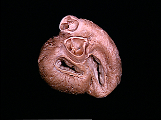
Ventricular myocardium; cardiac valves and ostia, basal view
Image #120-6
KEYWORDS: Heart, Left heart, Muscles and tendons, Right heart.
Creative Commons
Stanford holds the copyright to the David L. Bassett anatomical images and has assigned Creative Commons license Attribution-Share Alike 4.0 International to all of the images.

For additional information regarding use and permissions, please contact the Medical History Center.


Detailed dissection of heart
Ventricular myocardium; cardiac valves and ostia, basal view
The atria have been cut off close to their attachments to the atrioventricular valve rings.
- Pulmonary trunk
- Right semilunar cusp
- Anterior semilunar cusp
- Left semilunar cusp (together 2-4 make up the pulmonary valve
- Nodule of valve
- Left coronary artery (at origin from aortic sinus)
- Ascending aorta
- Left fibrous trigone
- Left ventricle
- Left atrioventricular opening
- Anterior cusp of mitral valve
- Posterior cusp of mitral valve
- Myocardium left atrium (cut across)
- Right fibrous trigone
- Atrioventricular bundle (bundle of His)
- Posterior interventricular sulcus
- Right atrioventricular opening
- Conus arteriosus
- Left semilunar cusp (aortic)
- Upper pointer: Right semilunar cusp Lower pointer: posterior semilunar cusp (of aortic valve)
- Right coronary artery (at origin from aortic sinus)
- Atrioventricular septum
- Anterior semilunar cusp (pulmonary)
- Septal (medial) cusp of tricuspid valve
- Posterior cusp tricuspid valve
- Right ventricle
- Myocardium right atrium (cut across)