
Bassett Collection of Stereoscopic Images of Human Anatomy
Exploration of the brain from its superior aspect
Systems of fibers related to head of caudate nucleus and putamen
Image #12-7
KEYWORDS: Brain, Telencephalon.
Creative Commons
Stanford holds the copyright to the David L. Bassett anatomical images and has assigned Creative Commons license Attribution-Share Alike 4.0 International to all of the images.

For additional information regarding use and permissions, please contact the Medical History Center.

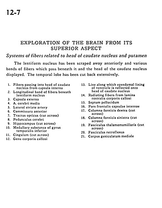
Exploration of the brain from its superior aspect
Systems of fibers related to head of caudate nucleus and putamen
The lentiform nucleus has been scraped away anteriorly and various bands of fibers which pass beneath it and the head of the caudate nucleus displayed. The temporal lobe has been cut back extensively.
- Fibers passing into head of caudate nucleus from internal capsule
- Longitudinal band of fibers beneath lentiform nucleus
- External capsule
- Middle cerebral artery
- Lateral striate artery
- Interior commissure
- Optic tract (cut across)
- Cerebral peduncle
- Hippocampus (cut across)
- Medullary substance of inferior temporal gyrus
- Cingulum (cut across)
- Genu corpus callosum
- Line along which ependymal lining of ventricle is reflected onto head of caudate nucleus
- Radiating fibers from rostral lamina corpus callosum
- Septum pellucidum
- Frontal part internal capsule
- Right column fornix (cut across)
- Left column fornix (cut across)
- Mamillothalamic tract (cut across)
- Fasciculus retroflexus
- Medial geniculate body