
Bassett Collection of Stereoscopic Images of Human Anatomy
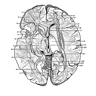
Exploration of the brain from its superior aspect
General orientation view of dissection of diencephalon, internal capsule and lentiform nucleus
Image #12-2
KEYWORDS: Brain, Diencephalon, Telencephalon, Overview.
Creative Commons
Stanford holds the copyright to the David L. Bassett anatomical images and has assigned Creative Commons license Attribution-Share Alike 4.0 International to all of the images.

For additional information regarding use and permissions, please contact the Medical History Center.

Exploration of the brain from its superior aspect
General orientation view of dissection of diencephalon, internal capsule and lentiform nucleus
The lentiform nucleus and internal capsule have been further cut away posteriorly. The geniculate bodies are sectioned horizontally. Relations of these structures to major landmarks of the brain are visible.
- Medullary substance of superior frontal gyrus
- Medullary substance of medial frontal gyrus
- Medullary substance of inferior frontal gyrus
- Lateral fissure
- Internal capsule
- Lentiform nucleus
- Cerebral peduncle (cut across)
- Lateral geniculate body
- Inferior horn of lateral ventricle
- Posterior horn lateral ventricle
- Lingual gyrus (cut across)
- Longitudinal fissure (cerebral)
- Cingulate gyrus
- Genu corpus callosum
- Inferior frontal sulcus
- Head of caudate nucleus
- Septum pellucidum
- Thalamus
- Third ventricle
- Lateral fissure
- Mesencephalon
- Cerebellum
- Superior temporal sulcus
- Surface of lingual gyrus facing calcarine fissure