Bassett Collection of Stereoscopic Images of Human Anatomy
Dissection of pericardium and heart in situ
Latex cast of cavities of left atrium and left ventricle; transverse sinus of pericardium
Image #118-4
KEYWORDS: Heart, Left heart, Pericardial sac.
Creative Commons
Stanford holds the copyright to the David L. Bassett anatomical images and has assigned Creative Commons license Attribution-Share Alike 4.0 International to all of the images.

For additional information regarding use and permissions, please contact the Medical History Center.


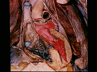
Dissection of pericardium and heart in situ
Latex cast of cavities of left atrium and left ventricle; transverse sinus of pericardium
The aorta and pulmonary trunk have been cut off to expose the transverse sinus of the pericardial cavity (3) and the left atrium. The anterior wall of the left atrium and the anterior cusp of the mitral valve have been resected to uncover the red latex cast which fills the left atrium and is continuous through the atrioventricular ostium with the cast of the left ventricle.
- Superior vena cava
- Ascending aorta
- Upper pointer: Transverse pericardial sinus Lower pointer: Wall of left atrium (cut across)
- Sinus of vena cava
- Membranous part interventricular septum
- Muscular part interventricular septum
- Blue latex within coronary sinus
- Pulmonary trunk
- Left auricle
- Red latex cast in left atrium (position of anterior segment of fibrous mitral valve ring indicated on drawing by dotted line below pointer)
- Latex cast within left atrioventricular opening
- Latex cast within left ventricle
- Anterior papillary muscle