
Bassett Collection of Stereoscopic Images of Human Anatomy
Dissection of pericardium and heart in situ
Latex cast of cavity of left ventricle and origin of aorta
Image #117-7
KEYWORDS: Heart, Left heart, Pericardial sac, Vasculature.
Creative Commons
Stanford holds the copyright to the David L. Bassett anatomical images and has assigned Creative Commons license Attribution-Share Alike 4.0 International to all of the images.

For additional information regarding use and permissions, please contact the Medical History Center.
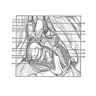
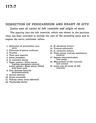
Dissection of pericardium and heart in situ
Latex cast of cavity of left ventricle and origin of aorta
The opening into the left ventricle, which was shown in the previous view, has been extended to include the root of the ascending aorta and to expose the aortic semilunar valves.
- Reflection of pericardium onto aorta
- Filaments of cardiac plexus
- Trachea
- Superior vena cava
- Ascending aorta
- Right coronary artery
- Upper pointer: Aortic valve (right semilunar cusp) Lower pointer: Aortic sinus (filled with latex cast)
- Right crus atrioventricular bundle
- Right atrium
- Crista terminalis
- Valve of inferior vena cava
- Right ventricle
- Left phrenic nerve
- Pulmonary trunk
- Left coronary artery
- Aortic valve (left semilunar cusp)
- Interventricular septum (cut away)
- Myocardium of left ventricle (cut across)
- Latex cast of cavity of left ventricle