
Bassett Collection of Stereoscopic Images of Human Anatomy
Dissection of pericardium and heart in situ
Ascending aorta; aortic arch and branches
Image #117-2
KEYWORDS: Heart, Left heart, Mediastinum, Pericardial sac, Right heart, Vasculature.
Creative Commons
Stanford holds the copyright to the David L. Bassett anatomical images and has assigned Creative Commons license Attribution-Share Alike 4.0 International to all of the images.

For additional information regarding use and permissions, please contact the Medical History Center.

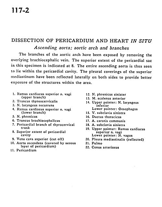
Dissection of pericardium and heart in situ
Ascending aorta; aortic arch and branches
The branches of the aortic arch have been exposed by removing the overlying brachiocephalic vein. The superior extent of the pericardial sac in this specimen is indicated at 8. The entire ascending aorta is thus seen to lie within the pericardial cavity. The pleural coverings of the superior mediastinum have been reflected laterally on both sides to provide better exposure of the structures within the area.
- Superior cardiac branch vagus nerve (upper branch)
- Thyrocervical trunk
- Recurrent laryngeal nerve
- Superior cardiac branch vagus nerve (lower branch)
- Phrenic nerve
- Brachiocephalic trunk
- Pericardial branch of thyrocervical trunk
- Superior extent of pericardial cavity
- Superior vena cava (cut off)
- Ascending aorta (covered by serous layer of pericardium)
- Pericardium
- Left phrenic nerve
- Anterior scalene muscle
- Upper pointer: Inferior laryngeal nerve Lower pointer: Esophagus
- Left subclavian vein
- Thoracic duct
- Common carotid artery
- Left subclavian artery
- Upper pointer: Superior cardiac branch vagus nerve Lower pointer: Vagus nerve
- Mediastinal pleura (reflected)
- Lung
- Conus arteriosus