
Bassett Collection of Stereoscopic Images of Human Anatomy
Dissection of pericardium and heart in situ
Heart and great vessels
Image #117-1
KEYWORDS: Heart, Left heart, Pericardial sac, Right heart, Vasculature.
Creative Commons
Stanford holds the copyright to the David L. Bassett anatomical images and has assigned Creative Commons license Attribution-Share Alike 4.0 International to all of the images.

For additional information regarding use and permissions, please contact the Medical History Center.

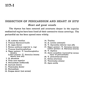
Dissection of pericardium and heart in situ
Heart and great vessels
The thymus has been removed and structures deeper in the superior mediastinal region have been freed of their connective tissue coverings. The pericardial sac has been opened more widely.
- Middle scalene muscle
- Thyrocervical trunk
- Vagus nerve right
- Superior cardiac branch vagus nerve
- Brachiocephalic trunk
- Upper pointer: Left brachiocephalic vein Lower pointer: Right internal thoracic vein (cut off)
- Phrenic nerve
- Superior vena cava
- Pericardium (reflected)
- Right auricle
- Right ventricle
- Pericardium
- Body of sternum (cut across)
- Trachea
- Common carotid artery
- Inferior thyroid vein (cut off)
- Upper pointer: Internal thoracic vein Lower pointer: Internal thoracic artery
- Thymic vein
- Ascending aorta (covered by serous layer of pericardium)
- Pericardiacophrenic vein
- Conus arteriosus
- Left ventricle
- Apex of heart