
Bassett Collection of Stereoscopic Images of Human Anatomy
Dissection of pericardium and heart in situ
Thymus and pericardium; superior sternopericardial ligament
Image #116-5
KEYWORDS: Pericardial sac, Thymus.
Creative Commons
Stanford holds the copyright to the David L. Bassett anatomical images and has assigned Creative Commons license Attribution-Share Alike 4.0 International to all of the images.

For additional information regarding use and permissions, please contact the Medical History Center.
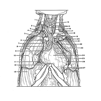

Dissection of pericardium and heart in situ
Thymus and pericardium; superior sternopericardial ligament
The internal thoracic arteries have been cut off (6, 21). The pleura has been cut away from the pericardium and the fascia which covered the thymus has been partially removed. A portion of this fascia (9) fuses firmly with the pericardium to form one of the sternopericardial ligaments. The sternal attachment of this ligament was in the area of origin of the sternothyroid muscles and was detached in the preparation of the dissection.
- Middle scalene muscle
- Anterior scalene muscle
- Right recurrent laryngeal nerve
- Pleura (cut to display apex of lung)
- Right brachiocephalic vein
- Upper pointer: Phrenic nerve Lower pointer: Internal thoracic artery (displaced posteriorly)
- Pericardiacophrenic artery
- Superior vena cava
- Superior sternopericardial ligament
- Right lung
- Mediastinal pleura (cut and reflected from pericardium)
- Body of sternum (cut across)
- Diaphragm
- Thyroid gland
- Common carotid arteries
- Vagus nerve left
- Thoracic duct
- Right subclavian artery
- Left brachiocephalic vein
- Rib I
- Internal thoracic artery
- Thymus (right lobe)
- Thymus (left lobe)
- Pericardium
- Left lung
- Mediastinal pleura (cut and reflected)
- Fibrous bands attaching pericardium to costal cartilages