
Bassett Collection of Stereoscopic Images of Human Anatomy
Dissection of breast and anterolateral thoracic wall
Seventh intercostal space.
Image #115-7
KEYWORDS: Bones joints cartilage, Muscles and tendons, Peripheral nervous system, Rib cage, Vasculature.
Creative Commons
Stanford holds the copyright to the David L. Bassett anatomical images and has assigned Creative Commons license Attribution-Share Alike 4.0 International to all of the images.

For additional information regarding use and permissions, please contact the Medical History Center.
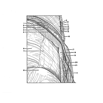
Dissection of breast and anterolateral thoracic wall
Seventh intercostal space.
The ribs above the eighth on the left side have been cut off along the mid-axillary line. The intercostal muscles of the seventh space have been retained and have been separated into their three layers (17, 18, 19). The middle layer, the internal intercostal muscle, becomes membranous in its posterior portion. The transition from muscle to membrane occurs just anterior to the cut end of the seventh rib (16). The membrane has been incompletely preserved in the seventh interspace. In the sixth interspace the membrane is clearly visible (14). The intercostal vessels and nerve have been separated slightly.
- Rib VIII
- Costal groove VII
- Posterior intercostal vein
- Posterior intercostal artery (a piece of this vessel several cms. in length has been removed laterally)
- Intercostal nerve VII
- Endothoracic fascia
- Costal pleura
- Diaphragm
- Posterior intercostal vein VI (cut off)
- Posterior intercostal artery VI (cut off)
- Intercostal nerve VI (cut off)
- Costal pleura
- Innermost intercostal muscle
- Internal intercostal membrane
- External intercostal muscle
- Rib VII (cut off)
- External intercostal muscle (reflected laterally)
- Innermost intercostal muscle (reflected inward)
- Internal intercostal muscle
- Muscular branch of intercostal nerve
- Lateral cutaneous branch intercostal nerve VII