Bassett Collection of Stereoscopic Images of Human Anatomy
Dissection of breast and anterolateral thoracic wall
Vessels and nerves of first and second left intercostal spaces; Internal thoracic vessels; sternal lymph nodes; sternocostal joint
Image #115-6
KEYWORDS: Bones joints cartilage, Lymphatics, Peripheral nervous system, Pleura, Rib cage, Vasculature.
Creative Commons
Stanford holds the copyright to the David L. Bassett anatomical images and has assigned Creative Commons license Attribution-Share Alike 4.0 International to all of the images.

For additional information regarding use and permissions, please contact the Medical History Center.
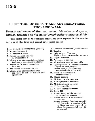
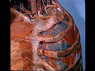
Dissection of breast and anterolateral thoracic wall
Vessels and nerves of first and second left intercostal spaces; Internal thoracic vessels; sternal lymph nodes; sternocostal joint
The costal part of the parietal pleura has been exposed in the anterior portions of the first and second intercostal spaces.
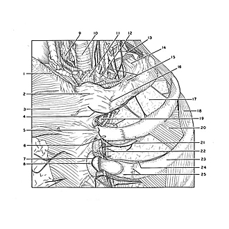
- Sternocleidomastoid muscle (cut off)
- Manubrium of sternum
- Pectoralis major muscle
- Anterior cutaneous branch intercostal nerve I
- Sternocostal ligament (pointer crosses sternal angle)
- Perforating branch internal thoracic artery
- Sternocostal joint III
- Sternocostal ligament (a delicate band in this specimen)
- Thyroid gland (right lobe)
- Trachea
- Left pointer: Thymus Right pointer: Common carotid artery
- Carotid sheath
- Left subclavian artery
- Anterior scalene muscle (cut off)
- Upper pointer: Internal thoracic artery (pointer near origin) Lower pointer: Costoclavicular ligament (cut off)
- Position of sternocostal joint I
- Costal pleura
- External intercostal muscle
- Sternal lymph nodes
- Internal intercostal muscle
- Intercostal nerve II
- Internal thoracic artery and vein
- Rib III
- Anterior intercostal branch internal thoracic artery
- Upper pointer: Costochondral junction Lower pointer: Costal cartilage III (note that a portion of the cartilage has been removed to allow the sternocostal joint to be opened)