Bassett Collection of Stereoscopic Images of Human Anatomy
Dissection of breast and anterolateral thoracic wall
Intercostal muscles, anterior view
Image #115-5
KEYWORDS: Bones joints cartilage, Breast, Muscles and tendons, Rib cage.
Creative Commons
Stanford holds the copyright to the David L. Bassett anatomical images and has assigned Creative Commons license Attribution-Share Alike 4.0 International to all of the images.

For additional information regarding use and permissions, please contact the Medical History Center.

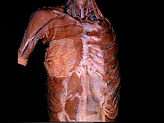
Dissection of breast and anterolateral thoracic wall
Intercostal muscles, anterior view
The external intercostal muscles have been removed from the first and second interspaces anteriorly to expose the internal intercostal muscles. A third, innermost layer of muscle has not been shown in this dissection. For a view of this layer reference should be made to 115-7.
- Clavicle
- Pectoralis major muscle
- Mammary body
- External abdominal oblique muscle
- Rectus abdominis muscle
- Vagus nerve left
- Trachea
- Internal jugular vein (at junction with subclavian vein)
- Common carotid artery
- Rib II
- Sternum
- Internal intercostal muscle
- External intercostal muscle
- External intercostal membrane
- Costochondral junction
- Sheath of rectus abdominis muscle
- Linea alba