Bassett Collection of Stereoscopic Images of Human Anatomy
Dissection of breast and anterolateral thoracic wall
External intercostal muscles viewed from left
Image #115-3
KEYWORDS: Bones joints cartilage, Muscles and tendons, Rib cage.
Creative Commons
Stanford holds the copyright to the David L. Bassett anatomical images and has assigned Creative Commons license Attribution-Share Alike 4.0 International to all of the images.

For additional information regarding use and permissions, please contact the Medical History Center.


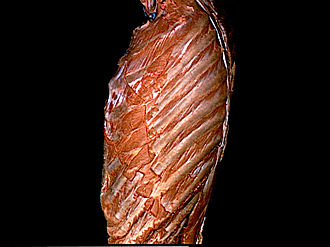
Dissection of breast and anterolateral thoracic wall
External intercostal muscles viewed from left
The left upper limb has been detached from the specimen.
- Rib I
- External intercostal muscle
- Pectoralis minor muscle (cut across near origin)
- Pectoralis major muscle (abdominal part, cut off near origin)
- External abdominal oblique muscle (cut off)
- Rib VIII (pointer on costochondral junction)
- Posterior scalene muscle
- Serratus anterior muscle (upper part, cut across)
- Trapezius muscle right
- Serratus anterior muscle middle part, cut across)
- Rib VI
- Serratus anterior muscle (lower part, cut across)
- Serratus posterior inferior muscle (cut across at insertions)
- Erector spinae muscle
- Rib XII