
Bassett Collection of Stereoscopic Images of Human Anatomy
Dissection of breast and anterolateral thoracic wall
Pectoral muscles
Image #115-2
KEYWORDS: Breast, Fascia and connective tissue, Muscles and tendons.
Creative Commons
Stanford holds the copyright to the David L. Bassett anatomical images and has assigned Creative Commons license Attribution-Share Alike 4.0 International to all of the images.

For additional information regarding use and permissions, please contact the Medical History Center.

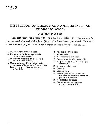
Dissection of breast and anterolateral thoracic wall
Pectoral muscles
The left pectoralis major (9) has been reflected. Its clavicular (2), sternocostal (3) and abdominal (4) origins have been preserved. The pectoralis minor (10) is covered by a layer of the clavipectoral fascia.
- Sternocleidomastoid muscle
- Clavicular part pectoralis major muscle (cut across)
- Sternocostal part of pectoralis major muscle (cut across)
- Upper pointer: Abdominal part pectoralis major muscle (cut across) Lower pointer: Sheath of rectus abdominis muscle
- Supraclavicular nerves
- Axillary vein
- Anterior thoracic nerve
- Remnant of pectoral fascia
- Pectoralis major muscle (reflected laterally)
- Pectoralis minor muscle
- Rib II
- Body of sternum
- Pectoral fascia (at former position of lateral border of pectoralis major)
- Serratus anterior muscle
- Lateral cutaneous branch intercostal nerve VII