Bassett Collection of Stereoscopic Images of Human Anatomy
Joints of right index finger
Metacarpophalangeal joint opened, medial view
Image #111-3
KEYWORDS: Fascia ligaments and tendons, Hand and fingers, Muscles and tendons.
Creative Commons
Stanford holds the copyright to the David L. Bassett anatomical images and has assigned Creative Commons license Attribution-Share Alike 4.0 International to all of the images.

For additional information regarding use and permissions, please contact the Medical History Center.
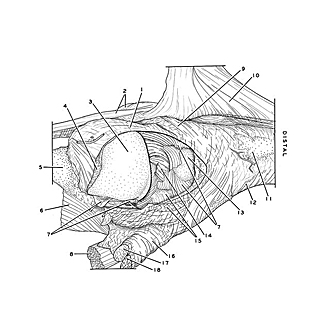
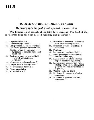
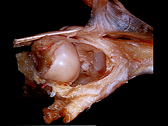
Joints of right index finger
Metacarpophalangeal joint opened, medial view
The ligaments and capsule of the joint have been cut. The head of the metacarpal bone has been rotated medially and proximally.
- Metacarpophalangeal joint capsule
- Left pointer: Extensor indicis muscle (tendon of insertion) Right pointer: Common extensor digitorum muscle (tendon of insertion)
- Head of metacarpal II (covered with articular cartilage)
- Collateral ligament (cut)
- Body of metacarpal II
- Dorsal interosseous muscle I
- Synovial fold
- Lumbrical muscle I
- Insertion of extensor tendons on base of proximal phalanx
- Extensor expansion (reflected dorsally)
- Phalanx I
- Ligament of digital sheath
- Base of phalanx (covered with articular cartilage)
- Collateral ligament (inner aspect of lateral ligament)
- Anterior accessory ligament (left pointer on cut edge, right pointer on articular surface of ligament)
- Tendon of digital sheath
- Flexor digitorum profundus muscle (tendon)
- Flexor digitorum superficialis (tendon)