Bassett Collection of Stereoscopic Images of Human Anatomy
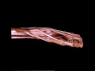
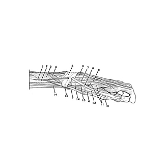
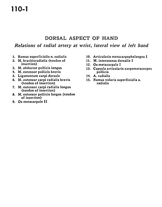
Dorsal aspect of hand
Relations of radial artery at wrist, lateral view of left hand
Image #110-1
KEYWORDS: Hand and fingers, Peripheral nervous system.
Creative Commons
Stanford holds the copyright to the David L. Bassett anatomical images and has assigned Creative Commons license Attribution-Share Alike 4.0 International to all of the images.

For additional information regarding use and permissions, please contact the Medical History Center.
Dorsal aspect of hand
Relations of radial artery at wrist, lateral view of left hand
- Superficial branch of radial nerve
- Brachioradialis muscle (tendon of insertion)
- Abductor pollicis longus muscle
- Extensor pollicis brevis muscle
- Dorsal carpal ligament
- Extensor carpi radialis brevis muscle (tendon of insertion)
- Extensor carpi radialis longus muscle (tendon of insertion)
- Extensor pollicis longus muscle (tendon of insertion)
- Metacarpal II
- Metacarpophalangeal joint I
- Dorsal interosseous muscle I
- Metacarpal I
- Carpometacarpal joint capsule of thumb
- Radial artery
- Superficial anterior branch radial artery