Bassett Collection of Stereoscopic Images of Human Anatomy
Exploration of the brain from its superior aspect
Associative systems of fibers within temporal lobe
Image #11-3
KEYWORDS: Brain, Telencephalon, Temporal lobe.
Creative Commons
Stanford holds the copyright to the David L. Bassett anatomical images and has assigned Creative Commons license Attribution-Share Alike 4.0 International to all of the images.

For additional information regarding use and permissions, please contact the Medical History Center.
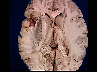
Exploration of the brain from its superior aspect
Associative systems of fibers within temporal lobe
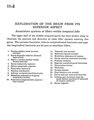
The upper half of the middle temporal gyrus has been broken away to illustrate the position and direction of some fiber systems entering this gyrus. The uncinate fasciculus, inferior occipitofrontal fasciculus and superior longitudinal fasciculus are all seen to contribute fibers.
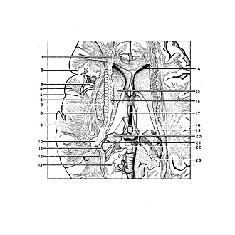
- Corona radiata (seen in cross section).
- Inferior frontal gyrus (frontal operculum)
- Branch middle cerebral artery within lateral fissure
- Superior temporal gyrus
- Superior temporal sulcus
- Uncinate fasciculus
- Lentiform nucleus
- Inferior occipitofrontal fasciculus
- Medullary substances of medial temporal gyrus
- Superior longitudinal fasciculus (cut across)
- Tapteum (cut across)
- External sagittal stratum
- Medullary substance of cuneus
- Anterior horn lateral ventricle
- Caudate nucleus
- Superior occipitofrontal fasciculus
- Third ventricle
- Thalamus
- Pineal body
- Fornix (crus) (cut across)
- Inferior horn of lateral ventricle
- Limbic lobe (just beneath former position of splenium of corpus callosum)
- Lingual gyrus