Bassett Collection of Stereoscopic Images of Human Anatomy
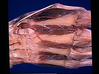
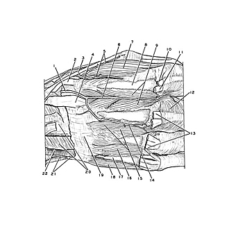
Dorsal aspect of hand
Dorsal interosseous muscles
Image #109-5
KEYWORDS: Hand and fingers, Vasculature.
Creative Commons
Stanford holds the copyright to the David L. Bassett anatomical images and has assigned Creative Commons license Attribution-Share Alike 4.0 International to all of the images.

For additional information regarding use and permissions, please contact the Medical History Center.
Dorsal aspect of hand
Dorsal interosseous muscles
The extensor tendons have been cut away and the dorsal interosseous fascia has been removed. A small part of the third dorsal interosseous muscle passes obliquely across the intermetacarpal space to insert on the shaft of the third metacarpal. The first dorsal interosseous muscle has a similar arrangement.
- Dorsal carpal branch radial artery
- Extensor carpi radialis longus muscle (tendon of insertion)
- Extensor pollicis longus muscle (tendon of insertion)
- Extensor carpi radialis brevis muscle (tendon of insertion)
- Branches of dorsal metacarpal artery I
- Dorsal metacarpal artery II
- Dorsal interosseous muscle I
- Metacarpal II
- Dorsal interosseous muscle II
- Common extensor digitorum muscle (tendon to index finger)
- Extensor indicis muscle (tendon of insertion)
- Transverse fibers of extensor expansion
- Common extensor digitorum muscle (tendons of insertion)
- Dorsal metacarpal artery III
- Dorsal interosseous muscle III (left pointer on muscle described in text above)
- Dorsal interosseous muscle IV
- Metacarpal V
- Abductor digiti minimi muscle
- Position of carpometacarpal articulation
- Left pointer: Dorsal carpal ligament Right pointer: Compartment for common extensor digitorum muscle and extensor indicis muscle
- Extensor carpi ulnaris muscle (tendon cut off within compartment)
- Compartment for extensor digiti minimi muscle