Bassett Collection of Stereoscopic Images of Human Anatomy
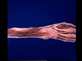
Dorsal aspect of right forearm
Deep layer of extensor muscles, general view
Image #108-1
KEYWORDS: Forearm, Vasculature.
Creative Commons
Stanford holds the copyright to the David L. Bassett anatomical images and has assigned Creative Commons license Attribution-Share Alike 4.0 International to all of the images.

For additional information regarding use and permissions, please contact the Medical History Center.
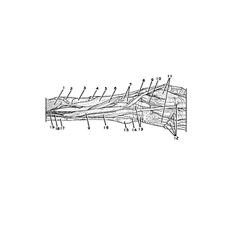

Dorsal aspect of right forearm
Deep layer of extensor muscles, general view
- Supinator muscle
- Pronator teres muscle (insertion)
- Radius
- Extensor carpi radialis longus and brevis muscles (tendons)
- Abductor pollicis longus muscle
- Extensor pollicis brevis muscle
- Left pointer: Extensor carpi radialis longus muscle (tendon) Right pointer: Extensor carpi radialis brevis muscle (tendon)
- Radial artery
- Extensor pollicis longus muscle
- Dorsal metacarpal artery II
- Dorsal interosseous muscles
- Common extensor digitorum muscle (tendons, cut off)
- Dorsal carpal ligament (compartments for extensor tendons opened)
- Compartment for extensor digiti minimi muscle
- Styloid process of ulna
- Extensor indicis muscle
- Intermuscular septum
- Dorsal interosseous artery
- Deep branch of radial nerve